


Osteoarthritis is a chronic disease in which the structure of the connective tissues of the musculoskeletal system is damaged. The disease is characterized by a progressive course, in the background of which the cartilage tissue is gradually destroyed. This pathology is diagnosed in many people over the age of 65, as one of the factors involved in the development of this condition is the natural aging process of the body.
Description of the disease
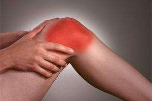
Post-traumatic, endocrine and inflammatory disease, excessive physical overload, or vice versa, inactivity can provoke the development of degenerative-dystrophic disease. The main signs of arthrosis are pain in the joint area with edema and limited activity.
Instrumental techniques - X-ray, arthroscopy, CT and MRI - are used to diagnose the disease. Conservative methods are used to treat stage 1 and 2 osteoarthritis - taking medications, physiotherapy, massage, and physiotherapy practices. If irreversible destructive changes have occurred in the joint tissue, surgery is needed - arthrodesis or endoprosthetics.
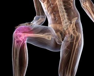
Pathogenesis
Osteoarthritis is characterized by marked changes in the structure of the connective tissue. Deformation erosions occur on the cartilage, resulting in the destruction of collagen fibers and proteoglycans, which contain protein (5-10%) and glycosaminoglycans (90-95%).
As a result, the stability of the collagen network decreases, metalloproteinase is released, and all proteins in the extracellular matrix are destroyed. The acceleration of death is due to an increase in the biosynthesis of collagenases and stromelysin.
As a general rule, when enzymes are present in normal amounts in the body, the levels of cytokines, small peptide information molecules, are kept in check. As arthrosis progresses, the concentration of this protein decreases, causing large amounts of enzymes that affect cartilage to be released.
As a result, distorted proteoglycans absorb water that they cannot retain. As a result, excess fluid penetrates the collagen fiber, which begins to "swell, " leading to a loss of strength and elasticity.
The qualitative and quantitative composition of synovial fluid also changes worse. In the background of arthrosis, a decrease in the concentration of hyaluronic acid is observed. The delivery of nutrients and oxygen to the hyaline cartilage tissue stops in the volume required to recover it. Softened foci are formed in the cartilage, followed by cracks and specific necrotic growths. Then the bare heads begin to expose, the microtrauma appearing in the background of the displacement relative to each other.
What provokes the development of the disease
Primary (idiopathic) arthrosis has not yet been established. Such a disease develops without affecting any factors, so doctors believe that the cause of such a problem lies in their predisposition to early destructive processes at the genetic level. The development of secondary arthrosis occurs as a complication of other joint diseases or as a background of injury.
The presence of the following may provoke the development of degenerative-dystrophic pathology:
- damage to joint tissue or injury near the connective tissue structure in the form of fracture, displacement, meniscus trauma, muscle and ligament tissue, partial rupture or complete rupture of tendon bone;
- congenital dysplastic disorder in the formation of joints;
- disorders of endocrine glands, metabolic disorders;
- rheumatism or rheumatic fever;
- polyarthritis, rheumatoid, reactive, metabolic, gouty or psoriatic arthritis;
- purulent arthritis due to the effects of streptococci, epidermal or Staphylococcus aureus;
- tuberculosis, brucellosis, chlamydia, gonorrhea, syphilis anywhere;
- degenerative-dystrophic pathologies such as osteochondritis dissecans.
Increased mobility of joint tissue, which is observed in the background of the production of special collagen fibers in the body, may contribute to the development of arthrosis.A similar phenomenon is observed in 10% of people on the planet, it is not considered a disease.Although there is a weakness in the tendon-ligament system behind hypermobility, which makes a person prone to injuries, especially in the ankle joint, in the form of ruptures of sprains and ligaments, dislocation.
In some cases, problems with hematopoietic function (such as the presence of hemophilia) can lead to arthrosis. In the background of hemarthrosis (bleeding into the joint cavity), the blood supply to the cartilage tissue deteriorates, causing it to collapse.
Among the predisposing factors, it is worth mentioning the presence of old age, the frequent load of joint tissue that exceeds the limits of its strength, excessive body weight, undergoing surgery, hypothermia.
Women at risk include citizens who are exposed to chemicals that live or are exposed to adverse environmental conditions during menopause. If the food does not contain enough vitamins and minerals, the conditions for the gradual destruction of hyaluronic cartilage will appear.
Symptoms

Arthrosis is dangerous because the first stage of development is asymptomatic. Manifestation of the clinical picture of the disease occurs over time, the primary symptoms appear with significant destruction of cartilage.At first, the patient feels mild pain syndrome without clear localization.Occurs after physical exertion - lifting heavy objects, sports training.
In some cases, the first sign a person notices the appearance of cracks and clicks as they bend or lengthen their joints. The patient notices that it is sometimes difficult to move. Although in the early stages of the development of arthrosis, mobility problems appear only in the morning and resolve rapidly.
As the pathology continues to develop, painful feelings begin to interfere at night, causing sleep function to be disrupted and chronic fatigue to appear. As the disease progresses to grade 2, the intensity of pain increases with changes in weather conditions, exacerbation of chronic diseases, underlying acute respiratory viral infections.
Physical activity decreases significantly. Mobility is inhibited by thinning of cartilage tissue and intentional restriction of the patient’s movement to prevent pain. This increases the load on the tissue of the opposite joint, which contributes to its further destruction.
Arthrosis has other features:
- pain that provokes the appearance of spasm in skeletal muscle and the development of muscle contracture (limited passive motor function of the joint);
- cracking in the joint tissue, clicks, cracks during constant movement, which practically occur when the bones move relative to each other;
- common painful muscle cramps;
- deformity of the joints leading to deterioration of posture and gait;
- pronounced deformity to the curvature of the joints with a significant decrease or complete absence of motor activity in the background of grade 3 arthrosis.
If arthrosis of the knee, ankle, or hip has developed until stage 3, one should use a reed or crutch when moving.
If you do not start treatment on time, the disease will progress, relapses will start to interfere regularly, and exacerbations will become more frequent over time. The stiffness of the morning hours does not go away for a long time, it gradually becomes constant.
When examining a person with stage 1 arthrosis, the doctor will notice only mild edema of the joint tissue with complete preservation of motor function. In stage 2 of the disease, pain and mild deformity occur on palpation. Bone thickenings develop near the synovial cavity.
Arthrosis is characterized by the development of synovitis - inflammation of the synovium in the hip, ankle, knee or shoulder joint. The main symptom of this disease is the formation of a rounded seal near the joint, when you press it, you can feel the liquid content moving. In acute synovitis, the temperature can rise to 37-38 degrees, headaches and digestive problems can occur.
Diagnostic Measures
The disease is diagnosed on the basis of the results of the examination with instrumental methods, clinical features, anamnestic data, patient complaints. In this case, the clinical examination of blood and urine is not very informative - all indicators remain within the normal range, unless the cause of arthrosis lies in metabolic problems.
When synovitis develops, the rate of erythrocyte sedimentation (up to 30 mm / h), leukocytes, and fibrinogen in the bloodstream increase.This indicates the presence of acute or chronic inflammation in the body.Biochemical and immunological parameters change with arthrosis of the secondary form.
The most informative way to detect degenerative-dystrophic diseases is by X-ray in two projections (lateral and straight).
X-rays show arthrosis as follows:
- There are no radiological signs in the initial phase.
- In the first stage, the pathology is presented as a vague, uneven stenosis of the joint cavity. The edges of the bone plates flatten slightly, initial osteophytes are formed (sometimes missing).
- In the second stage, the image shows an image in the form of a pronounced narrowing of the joint cavity that is 2-3 times the norm. Osteophytes are formed in large numbers, and the development of subchondral osteosclerosis is observed. Cyst-like enlightenments appear in the appendices.
- In the third stage, the image shows marked subchondral osteosclerosis and large marginal osteophytes. The joint space narrows significantly.
- In the fourth stage, coarse massive osteophytes are formed, the joint space is almost completely fused, and the bony appendages that make up the joint are deformed and compacted.
If the physician has any doubts about the diagnosis after examining the x-rays, the patient is prescribed a computed tomography scan. An MRI is performed to assess the condition of the connective tissue near the joint. The use of a contrast agent allows the dynamics of how tissues are supplied with blood to be monitored to determine the extent of inflammation in synovitis.
Arthrosis Treatment
It is currently impossible to completely cure arthrosis because there are no pharmacological agents that restore cartilage tissue.The main goal of treatment is to prevent the further development of the disease and to maintain the mobility of the joints.Therapy for arthrosis involves the use of long-term, complex, topical, and systemic medications.
Patients should not overload their joints, motor activity should be limited by orthopedic devices - orthoses, elastic bandages. Overweight people need to change their diet in order to lose weight over time and start losing weight.
To achieve stable remission, the patient should perform therapeutic gymnastic exercises every day. At first, you should perform it under the supervision of a professional, and in the future, you should exercise at home. In addition to physiotherapy, you can go to the pool, do yoga or go cycling.
In order to reduce the intensity of pain, the use of drugs belonging to different pharmacological groups is prescribed:
- Non-steroidal anti-inflammatory tablets, ointments, solutions for intravenous injection.
- Intra-articular injections of anesthetics with the addition of glucocorticosteroids.
- Muscle relaxants to relieve muscle cramps and contractures.
In addition, the treatment regimen for arthrosis includes the use of B vitamins, sedatives, antidepressants and sedatives if necessary. Chondroprotectors are mandatory in the form of a long course.The tools in this group help to partially restore cartilage.
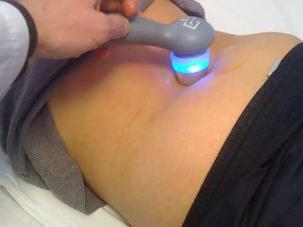
In order to increase the clinical activity of joint tissue, physiotherapy procedures should be performed - laser therapy, magnetotherapy, UHF.
Painful manifestations in the area of the joints should be the basis for immediate medical attention. Treatment in the early stages of the development of arthrosis makes it possible to stop the destructive processes in cartilage, to prevent disability and disability.