


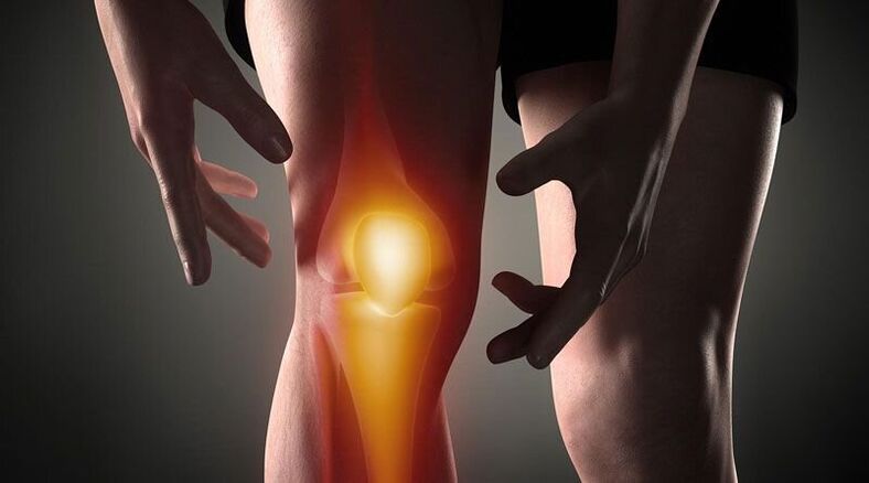
Knee pain- This is a sign of abnormal processes affecting the cartilage, bone or soft tissue structures of the femoral tibia and femoral patella joints. Arthralgia can be based on trauma, inflammatory and degenerative diseases of the joint apparatus and periarticular structures. Patients may complain of sharp, aching, burning, throbbing, and other pain that occurs at rest or when the foot is moved, supported, bent, and stretched in the knee. Diagnosis of causal pathology includes instrumental imaging methods (Rg, ultrasound, CT or MRI, arthroscopy), puncture of the joint capsule, biochemical and immunological analyzes. Until the diagnosis is clarified, rest, joint immobilization, NSAIDs, and analgesics are recommended.
Causes of knee pain
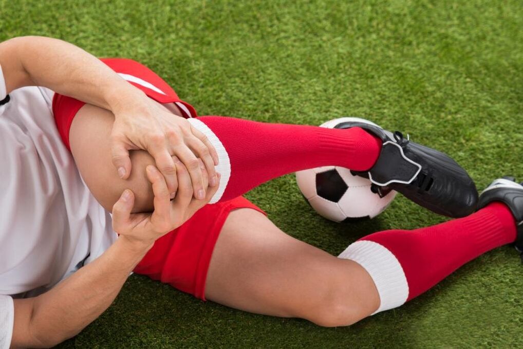
Traumatic injury
Usually the consequences of domestic traumas, often in athletes: runners, jumpers, athletes. It is caused by a fall, direct impact or twisting of the foot. It manifests itself in sharp pain at the time of the injury. In the future, the pain syndrome will become less pronounced, accompanied by increasing edema. Abrasions and bruises are possible. As the frequency increases, the following lesions are identified:
- Knee injury. . . Occurs on falling to the knee or direct impact. At first the pain is sharp, hot, sometimes burning, but tolerable, later - dull, sore, aggravated by movements. Bruising is possible. The footrest is retained. Sometimes the knee injury is complicated by hemarthrosis, in which case the joint gradually grows, becomes spherical, and the pain syndrome is accompanied by a feeling of pressure or cracking.
- Tear tape rupture. It can be detected in a non-physiological position after twisting, forcibly twisting, bending or over-tensioning the foot. Painful feelings are stronger than bruising; at the same time as the pain appears, one feels that something is torn (similar to the rupture of ordinary tissues). It is associated with significant restraint, support, limb twisting, and rapidly growing hemarthrosis.
- Intraarticular fractures. . . Collisions can be detected by falling and twisting the foot. In case of injury, the person feels very sharp, often unbearable sharp pain, sometimes a crack is heard. Patients with intra-articular fractures themselves describe their feelings as follows: "the pain is such that it darkens in your eyes, the world disappears, you don’t understand anything. " After that, the pains are milder but remain strong. Support is usually impossible, movement is almost completely restricted. Edema and hemarthrosis develop rapidly.
- Dislocation.The result of a blow or kneeling. As the kneecap moves, there is a sharp pain accompanied by a feeling of flexion of the leg and displacement of the knee. No movement is possible, the reference function can be saved. A pronounced deformation is seen on the anterior surface of the knee, which later smooths out due to increasing edema. Sometimes hemarthrosis is associated.
- Pathological fractures.They develop with minor injuries, osteoporosis, osteomyelitis, tuberculosis, and in the case of bone tumors, the consequences of a decrease in bone strength. The pains are aching, dull, reminiscent of pain syndrome with a bruise. Signs of abnormal fracture are limited or impossible to support the foot, a feeling of instability in the knee, sometimes deformity, bone cracking during movement.
- Damage to the meniscus.Meniscus tears are formed by twisting, collision, intense forced bending or knee stretching, sharp twists with fixed legs. At first, a person feels a special click and sharp shooting pain deep in the joint. Then the pain decreases somewhat, but it becomes diffuse, sometimes - burning, exploding, intensifying when they try to support and move it. Knee volume increases due to edema and hemarthrosis. Support becomes impossible, movements are sharply limited.
Inflammatory pathologies
They can be infectious and non-infectious (post-traumatic, toxic-allergic, metabolic, post-vaccination). The abundant blood supply to the synovial membrane and periarticular tissues promotes the rapid development of inflammation in response to direct and indirect effects, and the large number of nerve endings causes a pronounced pain response. The inflammatory process is often accompanied by arthritis (accumulation of aseptic fluid in the joint), which can cause pus to build up.
- Arthritis.Gonarthritis occurs after injuries, sometimes complicating infectious diseases, and is detected in rheumatic diseases. It can be acute or chronic. Knee pains are usually dull, sore, pressed, or pulled. At first, the pain is not intense and intermittent, intensifying in the evening or after training. Then the initial pains join, the intensity and duration of the pain syndrome increases. The joint swells, the skin blushes above it, and its temperature rises. In synovitis, the contours of the knee smooth out, there is a feeling of cracking. With suppuration, the severity of the pain increases sharply, they become twitching, causing sleep disturbances.
- Synovitis.It is not a disease in itself, complicating many acute and chronic pathologies of the joint. It develops within a few hours or days. Initially, the pain is insignificant or absent, a feeling of fullness prevails. The knee is spherical with a large amount of fluid, the skin is shiny. Movement is slightly restricted. When infected, the pain becomes pronounced, throbbing, twitching, intensified with the slightest movement and touch.
- Bursitis.Inflammation of the joint capsules in the patella and popliteal cavity usually occurs when the knee is overloaded and repeatedly injured (e. g. , with constant support of the knee). In bursitis, the pain is local, dull, not intense, appears in a certain position of the limb, after a characteristic load, decreases when the position of the foot changes, massaging the affected area. If the backpack is affected, painful sensations may occur as you ascend or descend the stairs. Sometimes minor local edema is defined. With the mucous membrane of the bursa, the pain becomes sharp, twitching, fried, with hyperemia, combined with edema of the affected area, symptoms of general poisoning.
- Tendinitis.It is usually seen in overweight men and athletes, affecting the patellar’s own ligament. At first, the pain syndrome occurs only during very intense exercise, then during normal sports exertion, and then during daily physical activity or at rest. The tendency associated with tendonitis is localized just below the knee, dull, pulling, as the disease progresses, sometimes paroxysmal, in some cases accompanied by mild redness and swelling, exacerbated by pressure. Movement is usually complete, less often slightly restricted. Tearing or tearing of the tape is possible due to a decrease in its strength.
- Lipoarthritis.Hoff's disease affects the layers of adipose tissue under the patella. It can be observed in case of permanent overload of the knee or it will be the result of an old injury. It affects athletes more often, older women. A person complains of dull aching pains combined with some limitation of extent. As the pathology worsens, the pain begins to disturb at night, a feeling of instability in the knee, and bending of the leg. You will hear a soft crack or squeak when you press the side of the kneecap.
Autoimmune processes
The diseases of this group are caused by the production of antibodies against normal cells of the body, the development of asymptomatic inflammation of the articular membrane and cartilage, the phenomena of vasculitis. Pathologies are in most cases chronic, prone to progression without treatment, and often cause disability.
- Rheumatoid arthritis.Defeat is usually bilateral. With minimal activity in the autoimmune process, pain is accompanied by weak or moderate, intermittent, pulling, compressive, morning stiffness. In the case of moderate activity, the patient complains of intermittent, prolonged, moderate-intensity aching, pushing, or tearing pains, not only during movement but also at rest. Stiffness for many hours, moderately recurrent arthritis. With high activity in rheumatoid arthritis, the pain is strong, diffuse, exhausting, wavy, and intensifies in the morning. Stiffness stabilizes, large amounts of fluid accumulate in the knee, and contractures develop over time.
- Systemic lupus erythematosus.Arthralgia is often symmetrical, although one joint may be affected. They can occur at any stage of the disease; in the recurrent course of SLE, they resemble rheumatoid arthritis. In the case of low activity of the process, the pain is short-term, not intense, local, sore, pulling. In severe cases, the pain syndrome progresses, the pain is wavy, interferes with nighttime sleep, prolongs, diffuses, increases movement, combined with arthritis, edema, hyperemia.
- Rheumatism.Joint pain is one of the first manifestations of rheumatic fever, occurring 5 to 15 days after acute infection, affecting multiple joints at once (usually in pairs). The pains are fairly short-lived but intense, migrating from one joint to another, ranging in character from pulling or pushing to burning or throbbing. The knees were swollen, hot, the skin reddened above them. Movement is severely restricted. After a few days, the severity of the pain decreases and the movements recover. In some patients, the residual effects persist for a long time in the form of moderate to mild dull pain.
- Reactive arthritis.It occurs more often 2 to 4 weeks after intestinal and urogenital infections, usually affecting one or two joints of the lower extremities, combined with urethritis, conjunctivitis. The development of reactive arthritis is preceded by increased urination, pain and a burning sensation in the urethra, tearing and cramps in the eyes. Knee pain is severe or moderate, constant, wavy, aching, pulling, twitching, with limited mobility, worsening of general condition, fever, severe swelling and redness of the affected area. Painful feelings and signs of inflammation persist for 3 months to 1 year and then gradually disappear.
Degenerative-dystrophic processes
They develop as a result of metabolic disorders in the structure of the joint and periarticular soft tissues. They are chronic and develop over many years. It is often accompanied by calcification, the formation of cysts and osteophytes, and deformation of the knee surface. With significant damage to the joint surfaces, they lead to marked impairment of movement and support function, become a cause of disability, and require the installation of an endoprosthesis.
- Osteoarthritis.It develops for no apparent reason or against the background of various injuries and diseases, especially in the elderly and middle-aged. The pain initially occurs with a weak, short-term, usually pulling or sore, prolonged load, disappears at rest, often accompanied by cracking. Gradually, the pain syndrome intensifies, the knees begin to hurt in "weather, " and movements are restricted at night. The distinguishing features of gonarthrosis are the onset of pain (until it "dissipates"), intermittent sharp cutting, burning, or shooting pain due to blockade. During the period of exacerbation, synovitis often occurs, in which the pain becomes constant, depressing, explosive.
- Meniscopathy. . . It is usually seen in athletes whose work puts a significant strain on the knee joint. It manifests as unilateral local deep pain inside the knee at the level of the joint gap, more often in the outer half of the knee. The pain intensifies as you move, passes at rest, and may be dull, depressing, or pulling. With progression, acute shooting pains occur when trying to move. On the anterolateral surface of the joint, a small painful formation can sometimes be felt in the projection of pain.
- Tendopathies. . . Tendons near the knee are affected. In the initial stage, they manifest as short-term local superficial pain at the peak of physical activity. This is followed by moderate and then mild painful feelings, limiting normal daily activities. The pain is pulling or aching, directly related to active movements, not noticeable during passive stretching and bending of the knee, sometimes accompanied by crackling or cracking. In the area of the lesion, the location of the greatest pain can be examined. Local signs of inflammation (edema, hyperemia, hyperthermia) are insignificant or absent.
- Osteochondropathy.Children and young people are more often affected, the duration of the disease is several years. They usually begin gradually with mild lameness or intermittent, non-intense, dull pains that are exacerbated by exertion and cease at rest. As osteochondropathy progresses, the pain becomes severe, persistent, depressing, burning, or burning, accompanied by severe lameness, limited mobility, and difficulty resting on the limb. After that, the pain gradually decreases and the support function is restored.
- Chondromatosis.It is usually diagnosed in older men, less often in infants. Joint chondromatosis is manifested by moderate dull wave-like pain, which often worsens at night and in the morning. Movement is limited, accompanied by crackling. Occasionally, blockages occur, characterized by sudden sharp pain, impossibility of movement, or severe restriction of movement. With the development of synovitis, the pain is explosive, combined with an increase in knee volume, swelling of the soft tissues, and a local rise in temperature.
Tumors and tumor-like formations
Pain syndrome can be caused by a cyst, benign, or malignant tumor that directly affects the joints or periarticular tissues. In addition, knee pain can serve as an alarming sign for hypertrophic arthropathy, paracancrotic polyarthritis - paraneoplastic syndromes characteristic of lung cancer, breast cancer, and other oncological processes.
- Baker's cyst.It represents the protrusion of the hernia of the popliteal fossa. In the initial stage, it manifests as discomfort or mild local pain in the back of the knee. Burning or shooting pain, numbness, or tingling in the area of the sole may occur in the background of an increased Baker cyst due to compression of nearby nerves. The symptoms are worse if we try to bend our knees as much as possible. In the popliteal cavity, a flexible, mildly painful tumor-like formation is sometimes felt.
- Benign tumors.These include chondromas, osteochondromas, non-sensitive fibroids, and other tumors. They are characterized by a prolonged asymptomatic or asymptomatic course, and may manifest as vague and intermittent local, non-intense pain. In the case of large neoplasms, a solid formation can be felt, sometimes synovitis develops.
- Malignant tumors.The most common malignancies affecting the joint area are synovial sarcoma, osteosarcoma, and chondrosarcoma. They are manifested by dull local, vague pains, sometimes with a certain circadian rhythm (worse night). The intensity of the pain increases, they become sharp, cut, burn or twitch, spread along the knee and adjacent tissues, with deformity, edema, synovitis, expansion of the saphenous veins, violation of the general condition, development of contracture. A painful tumor-like formation is defined by touch. At the start of the process, the pain is unbearable, exhausting, deprives you of sleep, and is not eliminated by non-narcotic painkillers.
Invasive surgeries and manipulations
Pain syndrome is triggered by damage to the knee tissue during invasive procedures. The severity of the pain depends directly on the trauma of the knee joint manipulations. With the penetration of pathogenic microbes into the joint area, the pain is caused by inflammatory changes.
- Manipulation.The most common procedure is stabbing. Post-puncture pain is short-lived, non-intense, rapidly attenuating, localized in the projection of the puncture, which is usually performed on the outer surface of the knee. After the biopsy, the pain may first be twitching, then dull, and disappear after a few days.
- Activities.After arthroscopy, the pain is moderate, at first quite acute, then dull, relieved after a few days or 1-2 weeks. After arthrotomy, the pain syndrome may persist for several weeks due to more intense, significant tissue damage. Usually, painkillers are prescribed to patients for the first 2-3 days after the procedure, after which the pain weakens and gradually disappears.
Psychosomatic conditions
Sometimes knee pain occurs in the absence of an organic basis (trauma, inflammation, destruction, etc. ) due to psychological factors. Such pain is believed to play a protective role as it helps reduce emotional stress by transforming experiences into physical feelings. The distinguishing features of such pains are their indeterminate nature, inconsistency, lack of visible changes, clear relationship to physical activity, and other objective provocative factors. Meteopathic arthralgias are observed in people who are sensitive to changes in atmospheric pressure.
In addition, irradiation of knee pain is possible in coxarthrosis, lumbar osteochondrosis, Perthes disease, fibromyalgia, sciatic nerve neuropathy. However, with these pathologies, pain syndromes with other localizations usually come to the fore. Other risk factors that increase the likelihood of knee injury and disease include being overweight, professional sports, hypovitaminosis, metabolic disorders, and old age. Hypothermia, stress, physical exertion, and eating disorders can be provoking factors in exacerbating chronic pain.
Survey
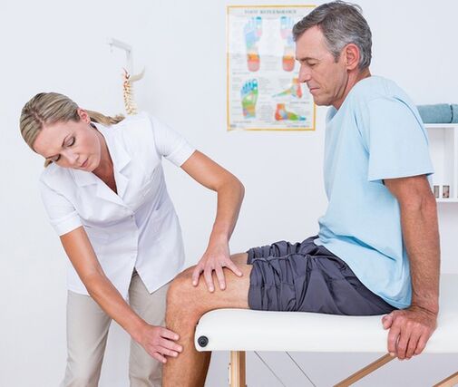
The diagnostic search algorithm is based on the nature and duration of the pain syndrome and the identification of simultaneous symptoms and events prior to the onset of knee pain. At the first visit of the doctor (traumatologist-orthopedist, surgeon, rheumatologist), a visual examination and palpation of the knee, assessment of the amount of active and passive movements is performed. Taking into account the received data, the following can be assigned to the patient in the future:
- Laboratory blood tests. . . A complete blood count helps to identify hematological changes (leukocytosis, increased ESR) characteristic of acute infectious and inflammatory processes, eosinophilia characteristic of allergic reactions. Biochemical and serological studies mostly provide information on autoimmune diseases characterized by the formation of specific acute phase proteins and immunoglobulins (CRP, rheumatoid factor, ASL-O, CEC, antibodies to DNA, etc. ).
- Radiography.The basic diagnostic method is X-ray of the knee joint in 2 projections. The presence of pathology is indicated by changes in the contours of the joint head and cavity, narrowing of the joint gap, changes in the thickness of the endplates, edge defects at the joint ends of the bones, osteolysis, and bone death. . In some diseases (meniscus trauma, Baker’s cyst), contrast arthrography shows the highest sensitivity.
- Arthrosonography. . . Knee ultrasound is a fast, inexpensive, affordable and highly informative diagnostic method. It allows the assessment of effusion and the presence of free bodies in the joint cavity, the identification of damage and pathological changes in periarticular soft tissues (signs of calcification, bleeding, etc. ). They help to distinguish the etiology of joint pain with great accuracy.
- CT and MRI. . . These are the methods of choice in arthropathy of any origin. They are used to assess the nature and extent of pathological lesions in more detail, and to identify signs of traumatic, inflammatory, and neoplastic lesions of bone structures and soft tissues. CT and MRI of the joints are commonly used with the limited information content of other instrumental examinations.
- Joint puncture. . . It is performed when there is a sign of accumulation of secretions or transudates in the joint capsule. As part of the differential diagnosis of inflammatory, degenerative and cancerous diseases, cytological, bacteriological or immunological examination of the synovial fluid is performed. To diagnose autoimmune damage of the knee joint, tuberculous arthritis, synovioma, it is extremely important to perform a biopsy of the joint membrane.
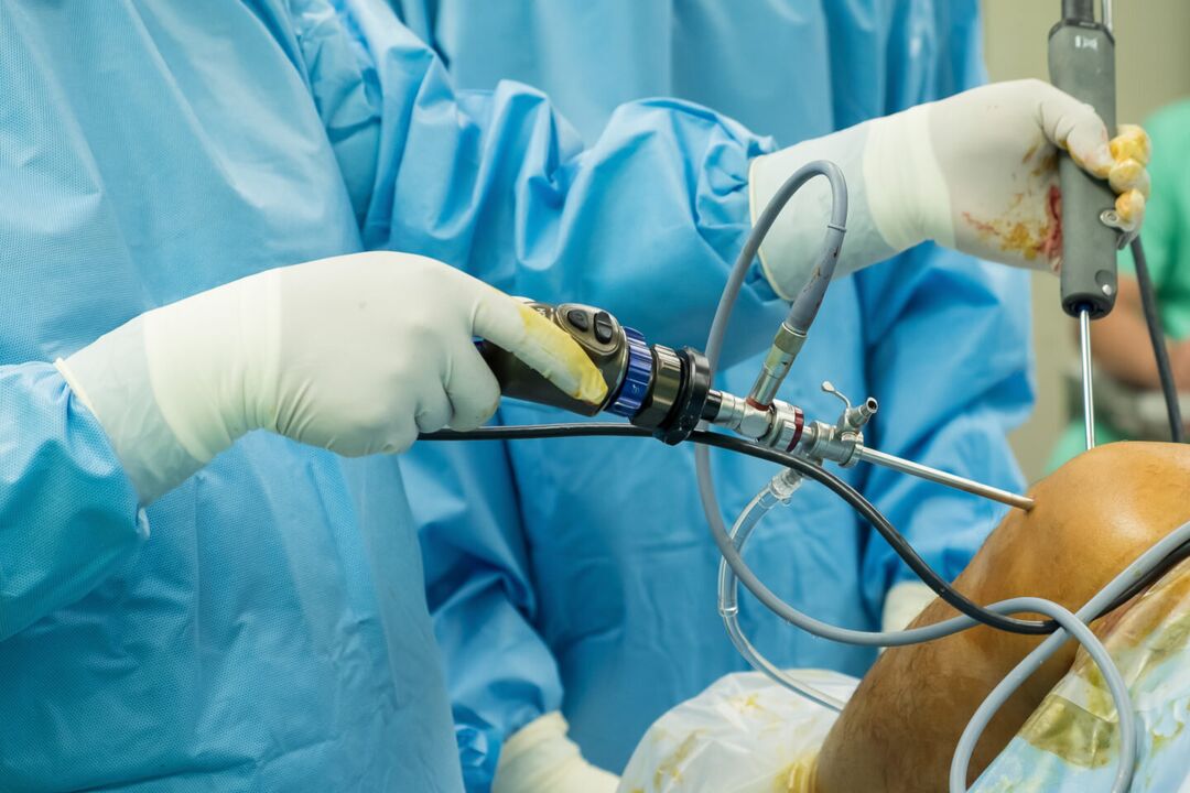
- Arthroscopy. . . The purpose of invasive endoscopic diagnostics may be biopsy sampling, clarification of the necessary diagnostic information during visual inspection of the joint elements. In some cases, diagnostic arthroscopy develops into therapy (atroscopic removal of intraarticular bodies, meniscectomy, autoplasty of ligaments, etc. ).
Symptomatic treatment
Treatment of the causes of knee pain is performed in a differentiated manner, taking into account the disease identified. At the same time, symptomatic care is an essential part of a comprehensive treatment process aimed at reducing discomfort and improving quality of life. Immediately after injury, it is recommended to apply a cold compress to the knee - this will help reduce the sensitivity to pain. Ethyl chloride has a local cooling and anesthetic effect. Resting your knee will always help reduce the pain. Movement should be restricted, giving the foot a position in which pain is minimal. A walking bandage is placed on the knee while walking, immobilization of the limb is possible with the help of plaster.
During the acute period of injury or illness, knee massage, use of a warming compress, and wearing high heels are strictly forbidden. The main groups of drugs used to symptomatically treat pain and inflammation are painkillers and NSAIDs in the form of ointments, tablets, and injections. The measures listed may only temporarily reduce pain, but do not eliminate the cause of arthralgia. Therefore, all cases of knee pain require professional diagnosis and treatment, and some conditions (fractures, dislocations, hemarthrosis) require emergency medical care. You should not delay a visit to the doctor if the pain is accompanied by a change in the shape of the knee (swelling, smoothing of contours, asymmetry), inability to perform bending-extensor movements, ballot knee ballotage, disturbance of knee support. the limb.