


Coxarthrosis (HJ) of the hip joint is a degenerative-dystrophic disease that affects cartilage and bone tissue. It can be called differently in medical articles: deforming coxarthrosis, hip joint DOA, osteoarthritis. All of these terms refer to the same pathology - arthrosis, but "coxarthrosis" is a narrower term that describes the defeat of the hip joint.
First, the cartilage suffers from arthrosis, and then the bones and surrounding structures — ligaments and muscles — are involved in the pathological process. If there are lesions in the bones, the prefix "osteo" is added to the word "arthrosis. "In advanced cases, the joint is deformed and there is talk of deforming arthrosis (osteoarthritis).
General features
Deforming arthrosis of the hip joint is the second most common after gonarthrosis of the knee. Due to the deep location of the hip joint, bone deformity will go unnoticed for a long time and only X-rays taken at later stages will show a change.
The development of the disease is influenced by a number of factors, including an inactive lifestyle, trauma, and metabolic disorders. Due to the peculiarities of modern life, in which there is often no room for physical education, arthrosis is affecting more and more people. In addition, the incidence of the peak falls in the middle age group - between 40 and 60 years.
Reference:coxarthrosis often affects women more than men.
Development mechanism
The femur is made up of two bones: the femur and the hip (pelvis). The head of the femur enters the acetabulum of the pelvis, which remains motionless during movement - walking, running. At the same time, the joint surface of the femur can move in several directions, providing flexion, stretching, abduction, adduction, and rotation of the thigh.
During physical activity, the femur moves freely in the acetabulum due to the cartilaginous tissue covering the joint surfaces. Hyaline cartilage is characterized by its strength, firmness and elasticity; it acts as a shock absorber and participates in the distribution of load during human movement.
Inside the joint is the synovial fluid - the synovium - which is essential for lubricating and nourishing cartilage. The entire joint is closed by a thick, thin capsule surrounded by strong muscles in the thighs and buttocks. These muscles also serve as shock absorbers to prevent injury to the hip joint.
The development of coxarthrosis begins with changes in the synovial fluid, which becomes more viscous and thicker. Due to the lack of moisture, the cartilage does not receive enough nutrients and begins to dry out, losing its smoothness, and cracks appear on it.
The bones can no longer move as freely as they used to and rub against each other, causing micro-damage to the cartilage. The pressure between the bones increases and the cartilage layer thins. As the pressure increases, the bones gradually deform and the local metabolic processes are disrupted. In the later stages, marked atrophy of the leg muscles occurs.
Cause
Deforming arthrosis of the hip joint may be primary or secondary. The cause of primary arthrosis cannot always be determined. Secondary arthrosis occurs in the background of existing diseases, namely:
- congenital hip dislocation or hip dysplasia;
- Perthes' disease (aseptic necrosis of the femoral head);
- coxarthritis of the hip joint of infectious, rheumatic or other origin;
- pelvic bone injuries - dislocations, fractures.
Hip dysplasia is a congenital disorder that sometimes does not occur clinically for a long time and can lead to dysplastic coxarthrosis in the future (aged 25-55 years).
Coxo arthrosis can be left, right and symmetrical. Concomitant diseases of the musculoskeletal system, especially osteochondrosis and gonarthrosis, are common in primary arthrosis.
There are also risk factors that contribute to the development of the disease:
- overweight and excessive loads that overload the joints;
- violation of blood circulation and metabolism;
- hormonal shifts;
- curvature of the spine, flat legs;
- old age;
- hypodynamics;
- heredity.
It should be noted that coxarthrosis itself is not inherited. However, certain characteristics of the metabolism or structure of connective tissues may create preconditions for the development of arthrosis in the child in the future.
Symptoms of coxarthrosis
The leading symptom of hip arthrosis is hip and groin pain of varying intensity. Stiffness and stiffness during movement, a decrease in muscle mass, a shortening of the affected limb, and a change in gait due to limping can also be observed.
Coxarthrosis usually progresses slowly, causing discomfort at first and mild pain after exercise. Over time, however, the pain intensifies and appears at rest.
A typical manifestation of the pathology is the difficulty of abducting the hip when a person cannot sit "in the stomach" on a chair. The presence and severity of signs of coxarthrosis depend on its extent, but pain syndrome is always present.
Three degrees or types of hip arthrosis differ in the severity of the injury and the accompanying symptoms:
- 1 degree. The thigh does not always hurt, but intermittently, especially after a long walk or standing. Pain syndrome is localized in the joint area, but can sometimes spread from the foot to the knee. Muscles with grade 1 coxarthrosis do not decrease in size, gait does not change, motor ability is fully maintained;
- Grade 2. The feeling of pain increases, not only after running or walking, but also at rest. The pain is more often concentrated in the thigh area, but can extend to the knee. In moments of heavy load, stepping on the injured limb is painful, so the patient begins to spare his leg, limping. The range of motion in the joint is reduced, it is especially difficult to move the foot to the side or to rotate the hip;
- 3 degrees. The pain perpetuates and does not ease even at night. The gait deteriorates noticeably, the independent movement becomes significantly more complicated, and the patient relies on a stick. The range of motion is sharply limited, the muscles of the buttocks and the entire leg, including the leg, atrophy.
- Due to muscle weakness, the pelvis bends forward and the affected leg shortens. To compensate for the difference in the length of the limbs, the patient tilts his body to the affected side while walking. This leads to a shift in the center of gravity and increased strain on the affected joint.
Osteoarthritis or osteoarthritis?
Arthritis is an inflammation of the joint that may be a stand-alone disease or may be the cause of systemic pathologies (such as rheumatism). In addition to the inflammatory response, symptoms of osteoarthritis (especially in the advanced stages) include limited mobility and joint deformity.
The focus of degenerative-dystrophic changes in arthrosis is the loss of cartilage tissue, which often leads to the appearance of inflammation. Therefore, arthrosis is sometimes referred to as arthrosis arthritis. And because arthrosis is almost always associated with joint deformity, the term "osteoarthritis" refers to it.
Reference:according to the International Classification of Diseases (ICD-10), osteoarthritis and osteoarthritis are variants of the same pathology.
Diagnosis of coxarthrosis
The diagnosis of "hip coxarthrosis" is based on the test, the patient's complaints and the test results. The most informative method is an x-ray: the images show both the degree of joint damage and the cause of the disease.
For example, in hip dysplasia, the acetabulum is flatter and sloping, and the cervico-diaphyseal angle (tilt of the femoral neck in the vertical plane) is larger than normal. Deformity of the part of the femur joint in the immediate vicinity is characteristic of Perthes' disease.
Grade 3 coxarthrosis is characterized by narrowing of the joint gap, dilation of the femoral head, and multiple bone outgrowths (osteophytes).
If the patient has had a fracture or dislocation, signs of trauma will also be visible on x-rays. If a detailed assessment of the condition of the bones and soft tissues is required, magnetic resonance imaging or computed tomography may be prescribed.
Differential diagnosis is made for the following diseases:
- gonarthrosis;
- osteochondrosis and radicular syndrome underlying it;
- trochanteritis (inflammation of the trochanterium of the thigh);
- ankylosing spondylitis;
- reactive arthritis.
The decrease in muscle volume that accompanies grades 2 and 3 of coxarthrosis can cause pain in the knee area. In addition, the knee often hurts even more than the hip joint itself. An X-ray is usually sufficient to confirm the diagnosis and rule out gonarthrosis.
Diseases of the spine - osteochondrosis and pinching of the nerve roots - are very similar to pain in coxarthrosis. However, this occurs unexpectedly after unsuccessful movement, a sharp turn, or weight lifting. The pain begins in the gluteal region and spreads to the back of the leg.
Radical syndrome is characterized by severe pain when a straight limb is lifted from a supine position. However, there are no difficulties in pulling the foot to the side as in coxarthrosis. It is worth noting that hip osteochondrosis and arthrosis are often diagnosed simultaneously, so a comprehensive examination is needed.
Trochanteritis or trochanteric bursitis develops rapidly, unlike arthrosis, which can progress slowly over years or even decades. Pain syndrome accumulates within a week or two while being quite intense. Trochanteritis is caused by trauma or excessive exercise. The movement is not restricted and the legs are not shortened.
Ankylosing spondylitis and reactive arthritis may also be associated with symptoms mimicking coxarthrosis. Such diseases are characterized by the onset of pain primarily at night. The hips can hurt quite a bit, but the pain subsides when walking and moving. In the morning, patients are worried about the stiffness that will disappear after a few hours.
Treatment of hip arthrosis
Coxarthrosis can be treated conservatively or surgically. The choice of treatment depends on the stage and nature of the pathological process. If grade 1 or 2 of the disease is diagnosed, it is treated with medication and physiotherapy. After alleviating the acute symptoms, therapeutic exercises and massages are added. If necessary, a special diet is prescribed.
The sooner coxarthrosis is detected and treated, the more favorable the prognosis. With the help of medications and therapeutic measures, you can significantly slow down the pathological process and improve your quality of life.
Non-steroidal anti-inflammatory drugs (NSAIDs) are used to relieve pain and inflammation. It should be noted that anesthesia is performed in the shortest possible time, as drugs in the NSAID class can negatively affect the digestive system and slow down the regeneration processes of cartilage tissue.
Chondroprotectors can be used to speed up cartilage recovery. However, these funds are only effective in the early stages of the disease, when hyaline cartilage is not completely destroyed. Chondroprotectors are prescribed in the form of tablets or intraarticular injections.
Vasodilators are used to improve the blood supply to the joint. Muscle relaxants are recommended for muscle cramps.
In case of persistent pain syndrome that is difficult to control with tablets, an injection is given into the hip joint. Corticosteroids are good for relieving inflammation and pain.
Medication therapy can be supplemented with topical agents - ointments and gels. They have no pronounced effect, but they help cope with muscle cramps and reduce pain.
Physiotherapy improves blood circulation and cartilage nutrition. For coxarthrosis, procedures such as shock wave therapy (SWT), magnetotherapy, infrared laser, ultrasound, and hydrogen sulfide baths have been shown to be excellent.
Operation
Treatment of stage 3 osteoarthritis can only be done surgically, as the joint is almost completely destroyed. Partial or complete arthroplasty is performed to restore the function of the hip joint.
Surgical treatment is used in advanced cases of arthrosis when conservative therapy is inert.
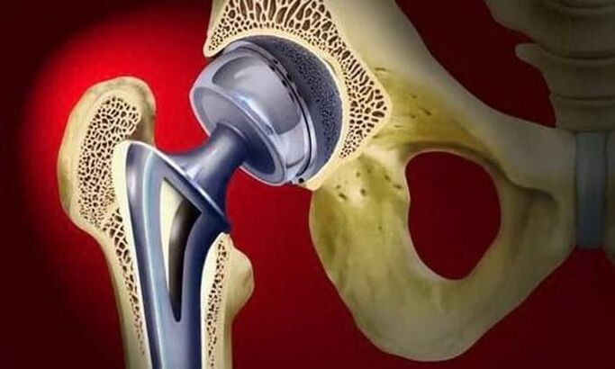
During a partial prosthesis, only the head of the femur is replaced with an artificial prosthesis. A complete prosthesis involves the replacement of the thigh head and acetabulum. Surgery is performed under general anesthesia, and in the vast majority of cases (approximately 95%), the function of the hip joint is completely restored.
During the rehabilitation period, the patient is prescribed antibiotics to prevent infectious complications. The seams are shown in Figures 10–12. days are removed and gym therapy begins. The attending physician will help the patient learn to walk and distribute the load correctly on the operated limb. Training is an important step in increasing muscle strength, endurance and flexibility.
Ability to work recovers on average 2-3 months after surgery, but in the elderly this period can be up to six months. Once rehabilitation is complete, patients can move, work, and even play sports. The life of the prosthesis is at least 15 years. A second surgery is performed to replace the worn prosthesis.
Effects
Timely and without proper treatment, coxarthrosis can not only significantly impair quality of life, but can also lead to disability and disability. Already in the second stage of arthrosis, the patient receives disability group 3.
When the affected limb is shortened by 7 cm or more, when a person moves only with improvised devices, a second group is assigned. Disability group 1 is given to patients with grade 3 coxarthrosis with complete loss of motor skills.
The indications for medical and social examination (MSC) are as follows:
- long course of arthrosis, more than three years, with regular exacerbations. Frequency of exacerbations at least three times every 12 months;
- underwent endoprosthesis surgery;
- severe musculoskeletal disorders of the limb.
Prophylaxis
The main measures to prevent coxarthrosis are diet (if you are overweight) and regular but moderate physical activity. It is very important to avoid damage to the pelvic region and hypothermia.
Swimming is beneficial in the presence of risk factors for the development of arthrosis as well as in all patients with a diagnosed disease. Sports such as running, jumping, soccer and tennis are not recommended.