


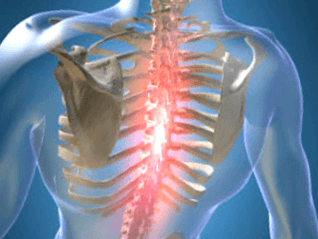
Unnatural changes in the cartilage and bones of the spine cause the development of the disease, which refers to the localization of M42 in accordance with the ICD-10 code and is called thoracic osteochondrosis. The middle part of the spine is subjected to less strain than the lumbar and cervical, but deformities are difficult to heal. The load is unevenly distributed due to the rounded configuration of the sternum, and osteophytes and other dysplastic manifestations appear.
Symptoms and signs
The disease occurs in the nucleus pulposus of the intervertebral disc, spreading to the fibrous fibrous ring and other parts of the vertebral segment that provide mobility to the spine. The changes are manifested in compression, reflex, or mixed neurological disorders and syndromes.
Pain is manifested by physical exertion. There are different feelings:
- mild prolonged pain in the chest region is called dorsalgia;
- sharp and sharp colic, causing difficulty in inhaling or exhaling, leading to immobility of the muscles - dorsago.
The symptoms and treatment of osteochondrosis of the thoracic spine depend on the degree of abrasion of the skeletal system and the stage of aging, which are common and local.
Symptoms include:
- damage to the peripheral processes of the nerves (neuralgia), characterized by painful attacks along the intercostal vessels;
- concentration of pain on the left side of the chest or a strong, shingles-like painful sensation;
- decreased mobility of the spine in the chest area;
- numbness in the arms and hands;
- decreased sexual function;
- the appearance of pain in the internal organs can cause heart, stomach, liver;
- lumbago in the neck, cheekbones and head, cough or lump in the throat;
- arrhythmia, tachycardia, fever.
Signs of osteochondrosis are disguised as manifestations of related diseases, so the symptoms are not clear. The nerves in the spinal cord focus around the spinal column as they constrict, sending signals to different parts of the body and organs.
Causes of osteochondrosis
There is no precise information on what factors deform intervertebral discs. A common cause of osteochondrosis is scoliosis or spinal curvature, which is more commonly recorded in childhood and adolescence.
The theory takes into account the following factors of vertebral deformity:
- dysontogenetic;
- hormonal;
- worth;
- functional;
- involutive;
- infectious;
- immunity;
- dysmetabolic;
- mechanical;
- hereditary.
Deterioration and aging of bones and cartilage occurs as a result of prior exposure to adverse conditions. Atrophic degenerations of the spine are determined by a genetic factor, and the disease with clinical symptoms develops in an exogenous and endogenous environment.
The consequence is in the form of complications in the work of the vertebrae, when the process of destruction of complex substances surpasses their synthesis. Aggravation occurs when the power supply to the disc is disrupted and the useful batteries are missing. The penetration of the elements and the dissimilation products decreases, the viability of the cells decreases, and the cell parts accumulate due to self-destruction. The production of complex proteins is reduced and collagen fibers are destroyed.
The mechanical effect on the annular bond formation is increased, the layered structure is disorganized, and the fibrous skeleton is torn. The disc is damaged by biomechanical factors and exercise, and its fixation is reduced. Due to the decrease in hydrostatic pressure, blood vessels and nerves can grow into the ring.
Diagnostic methods
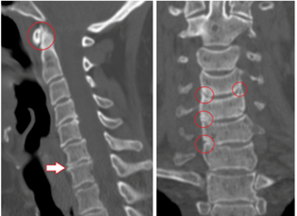
During recognition, radical, pain, reflex, myotonic, autonomic, and vascular factors can be identified. The best test method is difficult to identify because the diagnosis is made individually in each case.
The main methods are:
- X-ray diagnostics;
- CT scan;
- Magnetic resonance imaging.
The x-ray analyzes the condition of the spine, and the images are presented in oblique, lateral, and direct projections. Sometimes in a photo, a person bends, bends, or bends to the side.
Contrast radiography is divided into the following examinations:
- pneumomyelography - 20-40 ml of air is injected into the spinal canal;
- angiography - 10 ml of contrast is injected into the lumen of the vertebrae and 7-9 images are taken in 2-3 seconds;
- myelography - a coloration fluid is injected into the subarachnoid lumen and then the structure is scanned;
- discography - the stained material is injected directly into the plate for localized examination.
Computed tomography evaluates bone and tissue structure, the condition of blood vessels. The painless method takes three-dimensional images in a matter of minutes.
Benefits of CT:
- high detection speed;
- filtering "silent" areas during in-motion diagnostics;
- the possibility of multispiral angiography;
- recognition of long objects by making high-quality, small-thickness cuts.
MRI uses the magnetic field of a machine that builds up hydrogen atoms in the human body in parallel with the action. The particles indicate the response is recorded. The tomograph recognizes the waves and displays the result on the screen. There is no irradiation with MRI, the method is less dangerous but not recommended for pregnant women.
Treatment and prevention
Treatment of osteochondrosis is required in several stages, the complexity of which depends on the severity of the disease, contraindications and the resources of the body.
Mode:
- medicines and drugs;
- physiotherapy methods, exercises to remove the clamps, to relieve the patient's condition;
- operation.
There is a direction in kinesitherapy in which the treatment of spinal disorders in the form of hernia and spondylosis is possible with rehabilitation gymnastics. A method of postoperative recovery has also been developed.
Yoga practices help adult men, women, children overcome pain, warning that the most important thing is the psychological attitude.
Medicines
Medications are prescribed by a neurosurgeon or neuropathologist based on the card and medical history. Patients take medication in the hospital or at home, the point is to follow the instructions and not deviate from the intake.
Common medications:
- NSAIDs relieve pain, fever, and inflammation;
- muscle relaxants reduce skeletal muscle tone;
- hormones reduce neuralgic pain;
- vitamins B2, B6, B12, A and C are taken during remission and for simple prophylaxis;
- diuretics relieve swelling and release pinched radical nerves;
- neurometabolic stimulants improve metabolism in nerve tissue;
- Chondroprotectors restore cartilage in the vertebrae after injury.
Sometimes the patient is left without medication in the first stage of the onset of discomfort. Enough to exercise, use a massager.
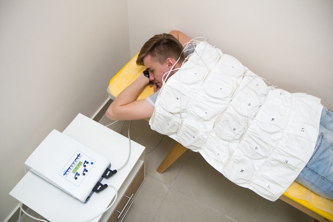
Physiotherapy
This type of exposure is used in combination with or separately from medication. In addition, bed rest is applied, heat is applied to the affected area. Folk recipes are used to relieve pain.
In a health care facility, physiotherapy involves the following procedures:
- ultrasound and phonophoresis;
- shock wave therapy;
- detensor blow;
- laser therapy;
- electrotherapy;
- magnetic waves;
- mud therapy and balneotherapy;
- massage.
Ultrasound refers to the effect of high-frequency waves on tissues, which reduces pain sensitivity. Painkillers and anti-inflammatory drugs are added during ultrophorophoresis to better reach the affected areas.
Shock wave therapy is used to transmit an acoustic wave to the painful area, to improve blood circulation and to speed up metabolism. Detensor therapy consists of stretching the spine using the patient's weight.
Laser therapy is based on the helium neon generation of lasers to activate bioelectric currents in nerve fibers. The laser acts on inflamed nerve roots in the paravertebral region along the chest region.
Electrotherapy improves nutrition and product metabolism in tissues, and pulse currents affect nerve endings. Low frequency waves relieve acute pain and are used as an initial relief.
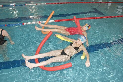
Magnetic therapy is used to relieve swelling, cramps and inflammation. A magnetic wave inductor is placed on the affected chest region. Balneotherapy and mud therapy consist of swimming in the pool, bathing, and contrast showers during treatment and healing. Metabolism is normalized, blood flow to the affected areas is accelerated, and pain and inflammation are reduced.
Therapeutic massage of osteochondrosis of the thoracic spine removes vacuum, puncture and lymph, improves blood microcirculation, tissue nutrition, tones muscles. Classes run by a knowledgeable professional if entrusting the spine to amateurs can have dangerous consequences. The massage is prescribed after the end of the acute phase, the first session should not exceed 10 minutes.
Operational treatment
The patient is advised to have surgery if medical treatment, massage and other procedures do not alleviate the condition.
The intervention is divided into 2 stages:
- eliminating the cause of severe pain (decompensation);
- stabilization of the spinal column.
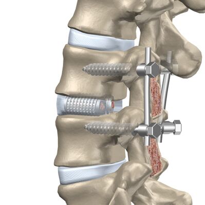
A retrospective approach involves a facetomy because beveled joints can depress the nerves. A foraminotomy is an extension of the radical channel through which the nerve leaves the vertebrae. Laminectomy removes the back of the vertebrae, which protects the lumen of the spine and pushes the brain due to deformity. Laminotomy involves enlarging the canal opening at the site of the spinal cord while removing a separate fragment of the back of the vertebrae.
Anterior surgery is performed when there is a protrusion (a bulge in the spine that protrudes toward the lumen) or a hernia that extends toward the canal.
The following methods are used for anterior decompression:
- discectomy - removal of an entire disc or a separate part of it;
- corpectomy - removal of a complete vertebrae and the adjacent plate and then implantation.
Discectomy and corpectomy lead to column destabilization and increase the risk of neurological disorders. Rigid fixation or fusion (fusion) of three vertebrae is used.
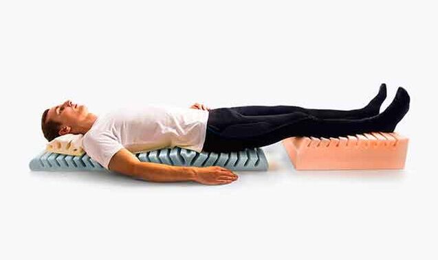
Prevention of chest osteochondrosis
Exacerbation of the disease reduces the ability to work and the quality of human life, so special attention is paid to prevention. As a result, vertebral degeneration appears later and disability can be avoided.
Methods of disease prevention:
- decreased physical activity of the spine;
- cannot stand still for long periods without changing the supporting limb, can lean on an impromptu object or wall;
- it is not recommended to sit at a table for a long time and when working with a computer you should take an active break, walk;
- mattresses and orthopedic headrests are selected for sleep;
- sudden turns and jumps should be avoided while running and walking, walking in low-heeled shock-absorbing shoes;
- Carry a weight not exceeding 10 kg, gradually lift it from a sitting position.
In the car, the backrests and headrests need to be fixed, while the driver’s seat needs to be rigid. Work cannot be performed in a reclining position, standing or sitting. Well-developed muscles support the skeleton, so they pay attention to feasible physical education and training.
Possible complications
The disease develops over a long period of time, sometimes the symptoms of pain do not appear immediately. Any degenerative change in the chest region leads to the appearance of pathologies.
Types of complications:
- cardiac pathology followed by myocardial infarction or angina pectoris;
- intercostal neuralgia or inflammation of the peripheral nerves with chest pain due to root compression;
- protrusion of intervertebral discs.
Complications occur with advanced forms of osteochondrosis, so early treatment in the early stages helps to avoid concomitant illnesses.